Dengue-Virus Infection: Lung Involvement, Clinical Implications, and Associated Human Leukocyte Antigens

Attapon Cheepsattayakorn1 and Ruangrong  Cheepsattayakorn2

110th  Zonal  Tuberculosis  and  Chest  Disease  Centre, Chiang  Mai, Thailand, 10th  Office  of  Disease  Prevention  and  Control, Department  of  Disease  Control, Ministry  of  Public  Health, Thailand

2 Department of Pathology, Faculty of Medicine, Chiang Mai University, Chiang Mai, Thailand

Academic Editor: Hirofumi Akari

Cite this Article as:

Attapon Cheepsattayakorn and Ruangrong Cheepsattayakorn (2014), " Dengue-Virus Infection: Lung Involvement, Clinical Implications, and Associated Human Leukocyte Antigens ", Journal of Respiratory Medicine Research and Treatment , Vol. 2014 (2014), Article ID 162245, DOI: 10.5171/2014.162245

Copyright © 2014. Attapon Cheepsattayakorn and Ruangrong Cheepsattayakorn. Distributed under Creative Commons CC-BY 3.0

Abstract

Four  known  dengue  virus  serotypes  have  been  identified, DENV-1, DENV-2, DENV-3, and  DENV-4.  About  50  million-infected  persons  occurs  each  year.  Adult  primary  infections  with  DENV-1  and  DENV-3  usually  result  in  dengue  fever  while  some  outbreak  with  DENV-2  infections  have  been  predominantly  subclinical.  Dengue  pulmonary  complications  include  pulmonary  infiltration, pleural  effusion, non-cardiogenic  pulmonary  edema  and  respiratory  failure  while  massive  hemoptysis  can  occur.  Several  human  leukocyte  antigens  class I  and  II  alleles  are  associated  with  the  development  of  dengue  disease.

Keywords: Dengue, Lung, HLA

Introduction
                  
This  disease  is  caused  by  dengue  virus (DENV)  that  belongs  to  the  family  Flaviviridae,genus  Flavivirus, and  is  transmitted  to  humans  by  Aedes  mosquitoes, mainly  Aedes  aegypti [Martina  et  al., 2009].  Four  serotypes  of  viruses  have  been  identified ; DENV-1, DENV-2, DENV- 3, and  DENV-4 [Martina  et  al., 2009].  An  estimated  50  million-infected  people  occur  each  year and  more  than  2.5  billion  people  are  being  at  risk  of  infection [Guha-Sapir  et  al., 2005], but  the  simultaneous  worldwide  distribution  of   the  risk  of  dengue  virus  infection  and  its  public  health burden  are  poorly  understood [Bhatt  et  al., 2013].  Epidemic  with  high  incidences  of  dengue hemorrhagic  fever  have  been  linked  to  primary  infection  with  DENV-1 followed  by  infection with  DENV-2  or  DENV-3,  whereas  it  indicated  that  the  longer  the  interval  between  primary and  secondary  infections, the  higher  the  risk  of  developing  severe  disease [Guzma”²n  et  al., 2003-Ong  et  al., 2007].  The  relationship  between  DENV-2  and  dengue  severity  is  controversial [Di”²az- Quijano  et  al., 2012].  However, adult  dengue  infections  are  frequently  accompanied  by  a  tendency for  severe  hemorrhage [UNICEF, UNDP, World  Bank, accessed  January  8, 2014]  and  can  be  life- threatening  when  infections  occur  in  patients  withchronic  diseases  such  as  asthma  and  diabetes  [Kouri  et  al., 1987-Lee  et  al., 2006].  The  aim  of  this  study  is  to  review  the  clinical  implications  and  associated  human  leukocyte  antigens  including  lung  pathological  mechanisms  and involvement  in  dengue  disease.      

Associated  Human  Leukocyte  Antigens, Lung  Involvement  and  Clinical  Implications

Lungs  of  dengue  cases, particularly  with  severe  disease,  present  with  mononuclear  inflammatory  infiltrates, hyperplasia  of  alveolar  macrophages, and  hyaline  membrane  formation  with  concomitant  hypertrophy  of  type  II  pneumocytes [Po”²voa  et  al., 2014].  Dengue  virus  antigens  with  virus  replication  are  also  identified  in  type  II  pneumocytes  and  pulmonary  vascular  endothelium [Po”²voa  et  al., 2014].  These  pulmonary  pathological  features  can  contribute  to pulmonary  edema, pulmonary  hemorrhage, adult  respiratory  distress  syndrome, and  pulmonary  tissue damages [Po”²voa  et  al., 2014].  Several  human  leukocyte  antigens (HLA)  class I  alleles, female  sex, AB  blood  group, a  single-nucleotide  polymorphism  in  the  tumor  necrosis  factor  gene, and  a  promoter  variant  of  the  DC-SIGN  receptor  gene  are  the  host  factors  that  increase  the  risk  of  severe  dengue  disease [Stephens  et  al., 2002-Kalayanarooj  et  al., 2007].  Notably, the  first  outbreak  in  the  Americas  occurred  in  1981, which  coincided  with  the  introduction  of  the  possibly  more  virulent  DENV-2  Southeast  Asian  genotype  whereas  the  less  virulent  indigenous  DENV-2  genotype  was  already  circulating  in  the  region [Kouri  et  al., 1987, Rico-Hesse  et  al., 1990-Rodriguez-Roche  et  al., 2005].  Age  has  been  demonstrated  to  influence  the  disease  outcome  following  a  secondary  infection  with  heterologous  DENV [Guzma”²n  et  al., 2002].  In  Asia, the  risk  of  severe  disease  is  greater  in  children  than  in  adults, in  contrast  to  the  Americas [Cologna  et  al., 2003, Leitmeyer  et  al., 1999].  Nevertheless, the  finding  that  dengue  hemorrhagic  fever  or  dengue  shock  syndrome  is  noted  primarily  in  a  relative  small  percentage  of  secondary  DENV  infections  and  to  a  much  lesser  extent  in  primary  infections  although  with  virulent  strains  it  indicates  that  host  factors  must  be  critical  determinants  of  severe  DENV  disease  development [Martina  et  al., 2009].  There  is  evidence  that  DENV  antigen  is  present  in  the  pulmonary  vascular  endothelium [Jessie  et  al., 2004], whereas  liver  is  the  organ  commonly  involved  in  human  DENV  infections  including  mouse  model [Paes  et  al., 2005, Seneviratne  et  al., 2006].

Glucose-6-phosphate  dehydrogenase  deficiency  which  is  highly  prevalence  among  the  African  population [Nkhoma  et  al., 2009]  can  cause  abnormal  cellular  redox, therefore  affecting  the  production  of  hydrogen  peroxide, superoxide, and  nitric  oxide  indicating  oxidative  stress [Wu  et  al., 2008].  Viral  proliferation  and  virulence  by  increasing  viral  receptors  on  target  cells  or  increasing  viral  particles  is  known  to  be  affected  by  oxidative  stress [Wu  et  al., 2008], therefore, glucose-6-phosphate  dehydrogenase  deficiency  may  contribute  to  the  increased  replication  of  DENV  in  monocytes [Nkhoma  et  al., 2009].  Several  HLA  class I  alleles (-A*01, -A*0207, -A*24, -B*07, -B*46, -B*51) [Stephens  et  al., 2002, Loke  et  al., 2002, Zivna  et  al., 2002]  and  HLA  class II  alleles (-DQ*1, -DR*1, -DR*4) [LaFleur  et  al., 2002, Polizel  et  al., 2004]  are  associated  with  the    development  of  dengue  hemorrhagic  fever.  Additionally, a  recent  study  demonstrated  that  there  was  significantly  higher  frequency  of  HLA-A*33  allele  in  dengue  fever  patients, HLA-A*0211  allele  in  dengue  hemorrhagic  fever  cases  compared  to  healthy  controls  and  dengue  fever  cases, respectively [Alagarasu  et  al., 2013].  The  frequency  of  HLA-B*18  and  HLA-Cw*07  alleles  were significantly  higher  in  DENV-infected  cases  compared  to  controls [Alagarasu  et  al., 2013].  The  combined  frequency  of  HLA-Cw*07  with  HLA-DRB1*07/*15  genotype  was  significantly  higher  in  dengue  hemorrhagic  fever  cases  as  compared  to  dengue  fever  cases  and  controls, but  the  frequency  of  combination  of  HLA-Cw*07  allele  without  HLA-DRB1*07  allele  was  significantly  higher  in  dengue  fever  cases  compared  to  controls [Alagarasu  et  al., 2013].  This  study  result  indicates  that  HLA-A*33  allele  may  be  associated  with  the  development  of  dengue  fever, whereas HLA-B*18  and  HLA-Cw*07  alleles  may  be  associated  with  symptomatic  dengue  infection  requiring  hospitalization [Alagarasu  et  al., 2013].  A  previous  study  demonstrated  that  HLA-A*0207  and  HLA-B*51  alleles  were  associated  with  dengue  hemorrhagic  fever  in  patients  having  secondary  DENV-1  or  DENV-2  infection  only   and  children  with  HLA-A*24  allele  were  more  likely  to  develop  dengue  hemorrhagic  fever [Malavige  et  al., 2004].  After  secondary  dengue  infections, HLA-B*44, -B*62, -B*76, and  -B*77  alleles  revealed  that  they  protect  against  development  of  clinical  disease [Malavige  et  al., 2004].

Rathakrishnan  and  colleagues  conducted  a  study  in  504  Chinese  and  Indian  Malaysian  populations, who  aged  14  and  above, which  demonstrated  that  HLA-A*24, HLA-A*33, and  HLA-B*57  alleles  were  positively  associated  with  patients  with  warning  signs  of  dengue  disease  or  severe  dengue  disease [Rathkrishnan  et  al., 2014].  HLA-A*03  allele  may  be  protective  in  both  Chinese  and  Indian  Malays, whereas  HLA-A*33  allele  may  be  a  predictive  marker  for  the  development  of  severe  dengue  disease [Rathkrishnan  et  al., 2014].  Cardozo  et  al  demonstrated  the  results  of  their  study  of  susceptibility  of  dengue  virus  serotype 3  among  95  patients  that  HLA-DQA1*05:01  and  HLA-DRB1*11  alleles  could  be  possible  resistance  factors  to  dengue  virus  serotype 3  infection, whereas  HLA-DQB1*06:11  and  HLA-DRB1*15  alleles  may  act  as  susceptible  factors [Cardozo  et  al., 2014].  Alencar  and  colleagues  conducted  a  study  of  a  cohort  of  dengue  patients  in  Brazil  and  demonstrated  that  HLA-B*44, HLA-B*50, HLA-DR*16  alleles  were  associated  with  increased  susceptibility  to  dengue  hemorrhagic  fever, particularly  serotype 3, whereas  HLA-B*07  and  HLA-DR*13  alleles  were  associated  with  resistance  to  secondary  dengue  infection  with  DENV-3 [Alencar  et  al., 2013].  Monteiro  et  al  conducted  a  retrospectively  case (dengue  hemorrhagic  fever)-control (dengue  fever)  study  among  Brazilians    during  2002-2008  and  revealed  that  HLA-A*01  allele  was  associated  with  increased  susceptibility  to  dengue  hemorrhagic  fever, whereas  HLA-A*31  allele  was  associated  with  resistance  to  the  development  of  dengue  hemorrhagic  fever [Monteiro  et  al., 2012].  Brown  and  colleagues  conducted  a  study  among  Jamaicans  and  demonstrated  that  HLA-A*24  and  HLA-DRbeta5*01/02  alleles  were  associated  with  increased  susceptibility  to  dengue  infection, whereas  HLA-A*23, HLA-CW*04, HLA-DQbeta*02, HLA-DQbeta*03, and  HLA-DQbeta*06  alleles  were  associated  with  protection  to  dengue  infection [Brown  et  al., 2011].  A  previous  study  in  Sri  Lanka  demonstrated  that  HLA-A*31  allele  was  associated  with  dengue  shock  syndrome  during  secondary  dengue  infection, while  HLA-A*24  and  HLA-DRB1*12  alleles  were  associated  with  the  development  of  dengue  hemorrhagic  fever  during  primary  dengue  infection [Malavige  et  al., 2011].

Appanna  et  al’s  study  demonstrated  that  HLA-B*18  and  HLA–B*53  alleles  were  increased  in  patients  with  dengue  hemorrhagic  fever, whereas  HLA-A*03  allele  was  decreased [Appanna  et  al., 2010].  Falco”²n-Lezama  and  colleagues  conducted  a  study  among  Mexican  patients  with  dengue  fever (23)  and  dengue  hemorrhagic  fever (16)  in  comparison  to  34  healthy  controls, and  revealed  that  HLA-DQB1*0202  and  HLA-DQB1*0302  alleles  were  associated  with  the  development  of  dengue  fever  and  dengue  hemorrhagic  fever, respectively [Falco”²n-Lezama  et  al., 2009].  A  study  in  228  ethnic  Thais  with  dengue  fever  and  142  patients  with  dengue  hemorrhagic  fever, which  was  further  classified  by  disease  severity  according  to  the  World  Health  Organization (WHO)  criteria, aged  3-14  years, demonstrated  that  HLA-B*48, HLA-B*57, and  HLA-DPB1*0501  alleles  were  associated  with  the  development  of  secondary  dengue  hemorrhagic  fever [Vejbaesya  et  al., 2009].  Lan  and  colleagues  conducted  a  study  in  Vietnam  by  gathering  211  patients  with  dengue  hemorrhagic  fever  and  418  patients  with  dengue  shock  syndrome  during  2002-2005, and  revealed  that  HLA-A*24  allele  was  associated  with  the  development  of  dengue  hemorrhagic  fever  and  dengue  shock  syndrome, whereas  HLA-DRB1*0901  allele  had  protective  effect  against    the  dengue  shock  syndrome  caused  by  DENV-2 [Lan  et  al., 2008].  Sierra  et  al  demonstrated  that  HLA-DRB1  polymorphism  was  associated  with  protective  effect  against  the    development  of  dengue  hemorrhagic  fever [Sierra  et  al., 2007].

Clinical  findings  in  early  febrile  stage  include  fever, headache, malaise, rash, body  pain, and  later  develop  pleural  effusion [UNICEF, UNDP, World  Bank, accessed  January  8, 2014, Likitnukul  et  al., 2004], both  lower  lobes  infiltration [Likitnukul  et  al., 2004], bilateral  perihilar  edema [Ali  et  al., 2010], ascites, bleeding, thrombocytopenia (platelet   20%, and  clinical  warning  signs  such  as  restlessness, severe  and  continuous  abdominal  pain, persistent  vomiting  and  a  sudden  reduction  in  body  temperature  associated  with  profuse  perspiration, adynamia (vigor  or  loss  of  strength)  and  sometimes  fainting  which  can  be  indicative  of  shock  due  to  plasma  extravasation [UNICEF, UNDP, World  Bank, accessed  January  8, 2014].  Wang  and  colleagues  conducted  a  study  in  661  Taiwanese  patients  diagnosed  with  dengue  fever  according  to  the  clinical  presentations  and  laboratory  examination  results  and  revealed  that  pleural  effusion  was  the  most  common  chest  roentgenographic  presentations (31.4%  of  the  total  chest  roentgenograms  and  57.4%  of  the  abnormal  chest  roentgenograms), followed  by  pulmonary  infiltration  only (23.3%  of  the  total  chest  roentgenograms  and  42.6%  of  the  abnormal  chestroentgenograms), while  small  pleural  effusion (less  than  2  intercostal  spaces)  was  the  predominate  type  among  the  chest  roentgenograms  presented  with  pleural  effusion  and  in  all  abnormal  chest  roentgenograms [Wang  et  al., 2007].

Additionally, pulmonary  infiltration  only  and  small  pleural  effusion  were  the  major  presentations  in  dengue  hemorrhagic  fever  patients  with  abnormal  chest  roentgenographic  features [Wang  et  al., 2007].  A  previous  study  conducted  in  100  patients  in  Yemen  with  seropositivity  of  dengue  confirmed  by  reverse-transcriptase  polymerase-chain-reaction  method, demonstrate  the  chest  presentations  as  the  following : 1) adult  respiratory  distress  syndrome  in  patients  with  dengue  fever, dengue  hemorrhagic  fever, and  dengue  shock  syndrome  was  0%, 19.4%, and  53.3%, respectively; 2) pulmonary  hemorrhage  in  patients  with  dengue  fever, dengue  hemorrhagic  fever, and  dengue  shock  syndrome  was  0%, 21.4%, and  6.6%, respectively; 3) unilateral  pneumonitis  in  patients  with  dengue  fever, dengue  hemorrhagic  fever, and  dengue  shock  syndrome  was  0%, 0%, and  0%, respectively; 4) bilateral  pneumonitis  in  patients  with  dengue  fever, dengue  hemorrhagic  fever, and  dengue  shock  syndrome  was  0%, 9.5%, and  6.6%, respectively; 5) pleural  effusion  in  patients  with  dengue  fever, dengue  hemorrhagic  fever, and  dengue  shock  syndrome  was  0%, 7.14%, and  0%, respectively ; 6) more  than  one  presentation  in  patients  with  dengue  fever, dengue hemorrhagic  fever, and  dengue  shock  syndrome  was  0%, 38.1%, and  33.3%, respectively [Mohamed  et  al., 2013].

Asghar  et  al  conducted  a  study  of  pulmonary  manifestations  in  76  confirmed  dengue-hemorrhagic-fever  patients, age  ranking  from  14  to  80  years, and  demonstrated  that  right  pleural  effusion, bilateral  pleural  effusion, left  pleural  effusion, bilateral  pneumonia, right  sided  pneumonia, and  left  sided  pneumonia  was  17.1%, 13.2%, 1.3%, 3.9%, 1.3%, and  0%, respectively [Asghar  et  al., 2011].  A  study  of  chest  computerized  tomography (CT)  in  29  Brazilian  patients  who  fulfilled  the  World  Health  Organization  case  definition  by  diagnoses  of  dengue  fever (9  patients)  and  severe dengue  disease (20  patients)  showed  that  abnormal  CT  findings  were  identified  in  58.62% (12  with  severe  disease, 5  with  dengue  fever), pleural  effusion  in  55.17% (11  with  severe  disease, 5  with  dengue  fever, 13  with  bilateral  effusion, 3  with  right-sided  effusion), large  pleural  effusion  in  4  patients  with  severe  disease [Rodrigues  et  al., 2014].  Large  pleural  effusion  was  not  identified  in  patients  with  dengue  fever [Rodrigues  et  al., 2014].  Ground-glass  opacity  was  the  most  common  finding  of  lung  parenchymal  involvement  that  was  noted  in  8  patients (5  with  severe  disease, 3  with  dengue  fever)  and  followed  by  lung  consolidation (6  patients (4  with  severe  disease, 2  with  dengue  fever)) [Rodrigues  et  al., 2014].  Interlobar  septal  thickening  and  pulmonary  nodules  with  no  specific  distribution  were  detected  in  2  patients (1  with  severe  disease, 1  with  dengue  fever, and  2  with  severe  disease, respectively) [Rodrigues  et  al., 2014].  One  case  with  dengue  fever  and  another  case  with  severe  disease  demonstrated  mild  interlobar  septal  thickening  that  located  predominantly  in  the  upper  lobes  and  lower  lung  zone, respectively [Rodrigues  et  al., 2014].  Only  one  case  with  intermediately  severe  disease  demonstrated  peribronchovascular  interstitial  thickening  in  the  middle  and  lower  zones [Rodrigues  et  al., 2014].  No  patient  with dengue  fever  showed  pulmonary  nodules [Rodrigues  et  al., 2014].   There  was  no  specific  axial  distribution [Rodrigues  et  al., 2014].  The  chest  extent  of  disease  tended  to  be  greater  in  patients  with  severe  disease  than  in  those  with  dengue  fever, but  this  difference  was  not  statistically  significant [Rodrigues  et  al., 2014].  Transudative  pleural  effusions  that  were  mostly  detected  in  dengue  patients  are  largely  due  to  imbalances  in  oncotic  and  hydrostatic  pressures  in  the  thoracic  cavity  because  these  effusions  are  ultrafiltrates  of  plasma [Wang  et  al., 2007].  Generally, the  identification  of  a  transudative  pleural  effusion  indicates  that  the  pleural  membranes  are  not  diseased  and  that  pleural  fluid  accumulation  is  caused  by  systemic (non-pleural, non-lung)  factors  affecting  the  formation  and  absorption  of  pleural  fluid [Wang  et  al., 2007].  Dengue  disease  must  be  excluded  from  two  syndromes  related  to  hantavirus, hemorrhagic  fever  with  renal  syndrome (HFRS)  in  Eurasia  and  hantavirus  pulmonary  syndrome (HPS)  in  the  Americas [Duchin  et  al., 1994-Vapalahti  et  al., 2003].  HPS  is  typically  characterized  by  acute  noncardiogenic  pulmonary  edema  and  circulatory  shock, whereas  fever, hemorrhagia, and  acute  renal  failure  are  hallmark  findings  in  HFRS [Rasmuson  et  al., 2011].

Laboratory  diagnosis  of  DENV  infection  includes  virus  isolation, serodiagnostic  tests (MAC-ELISA, IgG  ELISA, IgG : IgM  ratio, neutralization  assay), nucleic  acid  amplification  tests (real-time  PCR, reverse-transcriptase  PCR, nucleic  acid-sequence  based  amplification  assay (NASBA)), and  antigen  detection (NS1  antigen  and  antibody  detection) [UNICEF, UNDP, World  Bank, accessed  January  8, 2014].  DENV  complications  include  massive  hemorrhage  or  hemoptysis, disseminated  intravascular  coagulation, non-cardiogenic  pulmonary  edema, respiratory  failure, and  finally  develop  multiple  organ  failure [UNICEF, UNDP, World  Bank, accessed  January  8, 2014].  The  chest  roentgenographic  presentations  are  significantly  correlated  with  the  laboratory  findings, particularly  white  blood  cell  counts, platelet  counts, activated  partial  thromboplastin  time, and  serum  alanine  aminotransferase  and  albumin  levels [Wang  et  al., 2007].

In  uncomplicated  dengue  cases, treatment  is  only  supportive, but  in  cases  with  prolonged  or  recurrent  dengue  shock, intravenous  fluids  should  be  administered  carefully  according  to  dosage  and  age  to  prevent  pulmonary  edema [UNICEF, UNDP, World  Bank, accessed  January  8, 2014].  DENV  control  and  prevention  strategies  include  vector  control  and  vaccine  development [UNICEF, UNDP, World  Bank, accessed  January  8, 2014].  Current  approaches  to  vaccine  development  involve  using  deoxyribonucleic  acid  vaccine, chimeric  viruses  using  yellow  fever  vaccine, subunit  vaccine, inactivated  viruses, attenuated  viruses, and  attenuated  dengue  viruses  as  backbones[Guirakhoo  et  al., 2006-Edelman  et  al., 2003].  An  Acambis/Sanofi  Pasteur  yellow  fever-dengue  chimeric  vaccine  is  in  advanced  Phase II  testing  in  children  in  Thailand [UNICEF, UNDP, World  Bank, accessed  January  8, 2014].  A  possible  licensed  vaccine  will  be  available  in  less  than  10  years [UNICEF, UNDP, World  Bank, accessed  January  8, 2014].

Conclusions

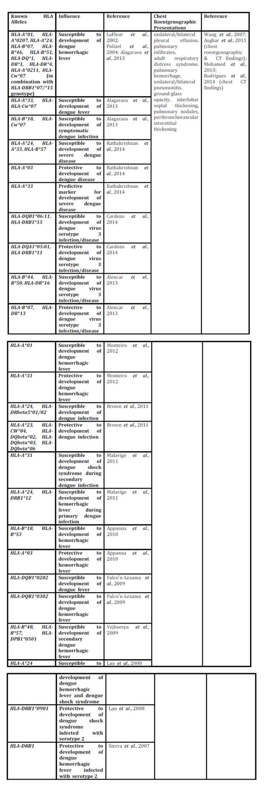
Presently, dengue  is  a  global  health  threat  and  is  tropically  endemic  or  epidemic.  Better  use  of  currently  available  measures  and  interventions  should  be  made  while  we  wait  for  novel  diagnostics, novel  vaccines, and  novel  antivirals.  Recently, several  partnerships  such  as  the  Asia-Pacific  Dengue  Prevention  Partnership  and  the  Innovative  Vector  Control  Consortium  have  come  into  existence  and  receive  funding  from  the  Bill  and  Melinda  Gates  Foundation, regional  Development  Banks  and  the  private  sector.  These  partnerships  are  collaborating  with  the  WHO  and  national  governmental  organizations  to  develop  novel  tools  and  strategies  to  improve  diagnostics, clinical  therapies, and  successful  novel  vaccines.  The  most  common  chest  presentation  is  pleural  effusion.  To  date, the  number  of  known  HLA  alleles  with  susceptible  effects  on  the  development  of  dengue  infection  or  severe  disease  are  more  than  the  number  of  known  HLA  alleles  with  protective  effects, thus, the  development  of  novel  protective  measures  against  dengue  virus  infection  are  urgently  needed  worldwide, particularly  in  the  tropical  regions.  The  summary  of  associated  HLA  alleles  and  chest  roentgenographic  presentations  in  dengue  disease  is  demonstrated  in  table 1.

Table 1:  Associated  Human  Leukocyte  Antigen  Alleles  and  Chest  Roentgenographic Presentations  in  Pulmonary  Dengue  Infection  or  Disease 

162245

References

1.Martina,  B.E.E., Koraka,  P. and  Osterhaus,  A.D.M. (2009), “ Dengue  virus  pathogenesis : an  integrated view, ”  Clinical  Microbiology  Reviews, 22 (4), 564-581.
PublisherGoogle Scholar

2.Guha-Sapir,  D.  and  Schimme,r  B (2005), “ Dengue  fever : new  paradigms  for  a  changing epidemiology, ”  Emerging  Themes  in  Epidemioogyl, 2 (1), 1.
Google Scholar

3.Guzma”²n,  M.G.  and  Kouri,  G (2003), “ Dengue  and  dengue  hemorrhagic  fever  in  Americas :lessons  and  challenges, ”  Journal  of  Clinical  Virology, 27 (1), 1-13. 
PublisherGoogle Scholar

 4. Bhatt,  S., Gething,  P.W., Brady,  O.J., Messina,  J.P., Farlow,  A.W., Moyes,  C.L., et  al (2013),
 “ The global  distribution  and  burden  of  dengue, ”  Nature, 496 (7446) : 504-507.
Google Scholar
 
5. Halstead,  S.B (2007), “ Dengue, ”  Lancet  370 (9599), 1644-1652.
PublisherGoogle Scholar

6. Ong,  A., Sandar,  M., Chen,  M.I.  and  Sin,  L.Y (2007), “ Fatal  dengue  hemorrhagic  fever  in  adults during  a  dengue  epidemic  in  Singapore, ”  International  Journal  of  Infectious  Diseases, 11 (3),263-267.
PublisherGoogle Scholar

7. Di”²az-Quijano  F.A.  and  Waldman  E.A. (2012), “ Factor  associated  with  dengue  mortality  in  LatinAmerica  and  Caribbean, 1995-2009 : an  ecological  study, ”  American  Journal  of  Tropical Medicine  and  Hygiene, 86 (2), 328-334.
PublisherGoogle Scholar

8. UNICEF, UNDP, World  Bank, WHO.  Evaluating  diagnostics-Dengue : a  continuing  global  threat.  (Retrieved  January  8, 2014),  http://www.nature.com/reviews/micro

9. Kouri,  G.P., Guzma”²n,  M.G.  and  Bravo,  J.R (1987), “ Why  dengue  hemorrhagic  fever  in  Cuba? 2.An  integral  analysis,  Transactions  of  the  Royal  Society  of  Tropical  Medicine  and  Hygiene, 81 (5), 821-823.

10. Halstead,  S.B., Nimanitaya,  S.  and  Cohen,  S.N (1970), “ Observations  related  to  pathogenesis  of dengue  hemorrhagic  fever : Relation  of  disease  severity  to  antibody  response  and  virus recovered, ” Yale  Journal  of  Biology  and  Medicine, 42 (5), 311-328.
Google Scholar

11. Lee,  M.S., Hwang,  K.P., Chen,  T.C., Lu,  P.L.  and  Chen,  T.P (2006), “ Clinical  characteristics  of dengue  and  dengue  hemorrhagic  fever  in  a  medical  center  of  southern  Taiwan  during  the  2002 epidemic, ”  Journal  of  Microbiology,  Immunology  and  Infection, 39 (2), 121-129.
Google Scholar

12. Po”²voa,  T.F., Alves,  Ada.M.B., Oliveira,  C.A.B., Nuovo,  G.J., Chagas,  V.L.A.  and  Paes,  M.V.P
 (2014), “ The  pathology  of   severe  dengue  in  multiple  organs  of  human  fatal  cases : histopathology, ultrastructure  and  replication, ”  PLoS  ONE, 9 (4), e83386.  DOI :10.1371/jpournal.pone.0083386
    
13. Stephens, H.A., Klaythong,  R., Sirikong,  M., Vaughn,  D.W., Green,  S., Kalayanarooj,  S., et  al (2002), “ HLA-A  and  HLA-B  allele  associations  with  secondary  dengue  virus  infections  correlate  with  disease  severity  and  the  infecting  viral  serotype  in  ethnic  Thais, ”  Tissue  Antigens, 60 (4), 309-318.
PublisherGoogle Scholar

14. LaFleur,  C., Granados,  J., Vargas-Alarcon,  G., Ruiz-Morales,  J., Villarreal-Garza,  C., Hiqueral,  L., et al (2002),
“ HLA-DR  antigen  frequencies  in  Mexican  patients  with  dengue  virus  infection : HLA- DR4  as  a  possible  genetic  resistance  factor  for  dengue  hemorrhagic  fever, ”  Human   Immunology,  63 (11), 1039-1044.
Google Scholar

 15. Loke,  H., Bethell,  D.B., Phuong,  C.X., Dung,  M., Schneider,  J., White,  N.J., et  al (2002), “ Strong HLA  class I-restricted  T-cell  responses  in  dengue  hemorrhagic  fever : a  double-edged  sword ? ” Journal  of    Infectious  Diseases, 184 (11), 1369-1373.

 16. Sakuntabhai,  A., Turbpaiboon,  C., Casade”²mont,  I., Chuansumrit,  A., Lowhoo,  T., Kajaste-Rudnitski, A., et  al (2005), “ A  variant  in  CD209  promoter  is  associated  with  severity  of  dengue  disease, ” Nature  Genetics, 37 (5), 507-513.
Publisher Google Scholar

17. Fernandez-Mastre,  M.T., Gendzekhadze,  K., Rivas-Vetencourt,  P.  and  Layrisse,  Z (2004), “ TNF-α-308A  allele, a  possible  severity  risk  factor  of  hemorrhagic  manifestation  in  dengue  fever  patients, ”  Tissue  Antigens, 64 (4), 469-472.
PublisherGoogle Scholar

18. Kalayanarooj,  S., Gibbons,  R.V., Vaughn,  D., Green,  S., Nisalak,  A., Jarman,  R.G., et  al (2007),  “ Blood  group  AB  is  associated  with  increased  risk  for  severe  dengue  disease  in  secondary infections, ”  Journal  of  Infectious  Diseases, 195 (7), 1014-1017.
PublisherGoogle Scholar

19. Rico-Hesse,  R (1990), “ Molecular  evolution  and  distribution  of  dengue  viruses  type 1  and  2  in nature, ”  Virology, 174 (2),  479-493.
PublisherGoogle Scholar

20. Rico-Hesse,  R., Harrison,  L.M., Salas,  R.A., Tovar,  D., Nisalak,  A., Ramos,  C., et  al (1997),
“Origins  of  dengue  type 2  viruses  associated  with  increased  pathogenicity  in  the  Americas, ” Virology, 230 (2), 244-251.
 Google Scholar

21. Rodriguez-Roche,  R.M., Alvarez,  M., Gritsun,  T., Halstead,  S., Kouri,  G., Gould,  E.A., et  al (2005),
“ Virus  evolution  during  a  severe  dengue  epidemic  in  Cuba, 1997, ”  Virology, 334 (2), 154-159.
Publisher Google Scholar

22. Guzma”²n,  M.G., Kouri,  G., Bravo,  J., Valdes,  L., Vazquez,  S.  and  Halstead,  S.B (2002), “  Effect  of age  on  outcome  of  secondary  dengue 2  infections, ”  International  Journal  of  Infectious  Diseases, 6 (2), 118-124.
PublisherGoogle Scholar

23. Cologna,  R.  and  Rico-Hesse,  R (2003), “ American  genotype  structures  decrease  dengue  virus output  from  human  monocytes  and  dendritic  cells, ”  Journal  of  Virology, 77 (7), 3929-3938.
PublisherGoogle Scholar

24. Leitmeyer,  K.C., Vaughn,  D.W., Watts,  D.M., Salas,  R., Villalobos,  I., Chacon,  de, et  al (1999),
“Dengue  virus  structural  differences  that  correlate  with  pathogenesis, ”  Journal  of  Virology, 73 (6),  4738-4747.
Google Scholar

25. Jessie,  K., Fong,  M.Y., Devi,  S., Lam,  S.K.  and  Wong.  K.T (2004), “ Localization  of  dengue  virus in  naturally  infected  human  tissues, immunohistochemistry  and  in  situ  hybridization, “Journal  of  Infectious  Diseases, 189 (8), 1411-1418.
PublisherGoogle Scholar

26. Paes,  M.V., Pinhao,  A.T., Barreto,  D.F., Costa,  S.M., Oliveira,  M.P., Nogueira,  A.C., et  al (2005),
“ Liver  injury  and  viremia  in  mice  infected  with  dengue-2  virus, ”  Virology, 338 (2), 236-246.
Google Scholar

27. Seneviratne,  S.L., Malavige,  G.N.  and  de  Silva,  H.J (2006), “ Pathogenesis  of  liver  involvement during  dengue  viral  infections, ”  Transactions  of  the  Royal  Society  of  Tropical  Medicine  and Hygiene, 100 (7), 608-614.
PublisherGoogle Scholar

28. Nkhoma,  E.T., Poole,  C., Vannappagari,  V., Hall,  S.A.  and  Beutler,  E (2009),
“ The  global prevalence  of  glucose-6-phosphate  dehydrogenase  deficiency : a  systematic  review  and  meta-analysis, ”  Blood  Cells  Molecules  and  Diseases, 42 (3), 267-278.
 Google Scholar

29. Wu,  Y.H., Tseng,  C.P., Cheng,  M.L., Ho,  H.Y., Shih,  S.R.  and  Chiu,  D.T (2008),
“ Glucose-6- phosphate  dehydrogenase  deficiency  enhances  human  coronavirus 229E  infection, ”  Journal  ofInfectious  Diseases,  197 (6), 812-816.
Google Scholar

30. Zivna,  I., Green,  S., Vaughn,  D.W., Kalayanarooj,  S., Stephens,  H.A., Chandanayingyong,  D., et  al
(2002), “  T-cell  responses  to  an  HLA-B*07-restricted  epitope  on  the  dengue  NS3  protein correlate  With  disease  severity, ”  Journal  of  Immunology, 168 (11), 5959-5965.
Google Scholar

31. Polizel,  J.R., Bueno,  D., Visentainer,  J.E., Sell,  A.M., Borelli,  S.D., Tsuneto,  L.T., et  al (2004),  “Association  of  human  leukocyte  antigen  DQ1  and  dengue  fever  in  a  white  Southern  Brazilian population, ”  Memo”²rias  do  Instituto  Oswaldo  Cruz, 99 (6), 559-562.
Publisher – Google Scholar

32. Alagarasu,  K., Mulay,  A.P., Sarikhani,  M., Rashmika,  D., Shah,  P.S.  and  Celilia,  D (2013), “  Profile of  human  leukocyte  antigen  class I  alleles  in  patients  with  dengue  infection  from  Western  India, ”  Human  Immunology, 74 (12), 1624-1628.
Publisher – Google Scholar

33. Malavige,  G.N., Fernando,  S., Fernando,  D.J.  and  Seneviratne,  S.L (2004), “ Dengue  viral  infection,  ”  Postgraduate  Medical  Journal, 80 (948), 588-601.
Publisher – Google Scholar

34. Rathakrishnan,  A., Klekamp,  B., Wang,  S.M., Komarasamy,  T.V., Natkunam,  S.K., Sanchez-Anguiano,  A, et  al (2014), “  Clinical  and  immunological  markers  of  dengue  progression  in  a  study  cohort  from  a  hyperendemic  area  in  Malaysia, ”  PLoS  ONE, 9 (3), e92021.  DOI : 10.1371/journal.pone.0092021
Publisher – Google Scholar

35. Cardozo,  D.M., Moliterno,  R.A., Sell,  A.M., Guelsin,  G.A.S., Beltrame,  L.M., Clemintino,  S.L., et  al (2014), “  Evidence  of  HLA-DQB1  contribution  to  susceptibility  of  dengue  serotype 3  in  dengue  patients  in  southern  Brazil, ”  Journal  of  Tropical  Medicine, Article  ID  968262, 6  pages.
Publisher – Google Scholar

36. Alencar,  L.X.E.de, Braga-Neto,  U.deM., Nascimento,  E.J.M.do, Cordeiro,  M.T., Silva,  A.M., Brito,  A.A.de, et  al (2013), “ HLA-B*44  is  associated  with  dengue  severity  caused  by  DENV-3  in  a  Brazilian  population, ”  Journal  of  Tropical  Medicine, Article  ID  648475, 11  pages.
Publisher – Google Scholar

37. Monteiro,  S.P.,Brasil,  P.E.A.A.do, Cabello,  G.M.K., Souza,  R.V.de, Brasil,  P., Georg,  I.,  et  al (2012),
“  HLA-A*01 allele : a  risk  factor  for  dengue  hemorrhagic  fever  in  Brazil’s  population, ”  Memo”²rias  do  Instituto  Oswaldo  Cruz, Rio  de  Janeiro, 107 (2), 224-230.
Publisher – Google Scholar

38. Brown,  M.G., Salas,  R.A., Vikers,  I.E., Heslop,  O.D.  and  Smikle,  M.F (2011), “  Dengue  HLA  associations  in  Jamaicans, ”  West  Indian  Medical  Journal, 60 (2), 126-131.
Publisher – Google Scholar

39. Malavige,  G.N., Rostron,  T., Rohanachandra,  L.T., Jayaratne,  S.D., Fernando,  N., Silva,  A.D.De, et  al (2011),
“  HLA  class I  and  class II  associations  in  dengue  viral  infections  in  a  Sri  Lankan  population, ”   PLoS  ONE, 6 (6), e20581.  DOI : 10.1371/journal.pone.0020581
Publisher – Google Scholar

40. Appanna,  R., Ponnampalavanar,  S., See,  L.L.C.  and  Sekaran,  S.D (2010), “  Susceptible  and  protective  HLA  class 1  allele  against  dengue  fever  and  dengue  hemorrhagic  fever  patients  in  a  Malaysian  population, ”  PLoS  ONE, 5 (9), e13029.  DOI : 10.1371/journal.pone.0013029
Publisher – Google Scholar

41. Falco”²n-Lezama,  J.A., Ramos,  C., Zuñiga,  J., Jua”²rez-Palma,  L., Rangel-Flores,  H., Garcia-Trejo,  A.R., et  al (2009), “  HLA  class I  and  II  polymorphisms  in  Mexican  Mestizo  patients  with  dengue  fever, ”  Acta  Tropica, 112 (2), 193-197.
Publisher – Google Scholar

 42. Vejbaesya,  S., Luangtrakool,  P., Luangtrakool,  K, Kalayanarooj,  S., Vaughn,  D.W., Endy,  T.P., et  al (2009),
 “ TNF  and  LTA  gene, allele, and  extended  HLA  haplotype  associations  with  severe  dengue  virus  infection  in  ethnic  Thais, ”  Journal  of  Infectious  Diseases,  199 (10), 1442-1448.
Publisher – Google Scholar

43. Lan,  N.T.P., Kikuchi,  M., Huong,  V.T.Q., Ha,  D.Q., Thuy,  T.T., Tham,  V.D., et  al (2008), “  Protective  and  enhancing  HLA  alleles, HLA-DRB1*0901  and  HLA-A*24, for  severe  forms  of  dengue  virus  infection, dengue  hemorrhagic  fever  and  dengue  shock  syndrome, ”  PLoS  Neglected  Tropical  Diseases, 2 (10), e304.  DOI : 10.1371/journal.pntd.0000304
Publisher – Google Scholar

44. Sierra,  B., Alegre,  R., Pe”²rez,  A.B., Garcia,  G., Sturn-Ramirez,  K., Obasanjo,  O., et  al (2007), “  HLA-A, -B, -C, and  -DRB1  allele  frequencies  in  Cuban  individuals  with  antecedents  of  dengue 2  disease : advantages  of  the  Cuban  population  for  HLA  studies  of  dengue  virus  infection, ”  Human  Immunology, 68 (6), 531-540. 
Publisher – Google Scholar
                                        
45. Likitnukul,  S., Prappal,  N., Pongpunlert,  W., Kingwatanakul,  P.  and  Poovorawan,  Y (2004), “  Dual  infections : dengue  hemorrhagic  fever  with  unusual  manifestations  and  mycoplasma  pneumonia  in  a  child, ”  Southeast  Asian  Journal  of  Tropical  Medicine  and  Public  Health, 35 (2), 399-402. 
Publisher – Google Scholar

46. Ali,  F., Saleem,  T., Khalid,  U., Mehwood,  S.F.  and  Jamil,  B (2010), “  Crimen-Congo  hemorrhagic fever  in  a  dengue-endemic  region : lessons  for  the  future, ”  Journal  of  Infection  in  Developin Countries, 4 (7), 459-463.
Publisher – Google Scholar

47. Wang,  C.C., Wu,  C.C., Liu,  J.W., Lin,  A.S., Liu,  S.F., et  al (2007), “ Chest  radiographic  presentation  in  patients  with  dengue  hemorrhagic  fever, ”  American  Journal  of  Tropical  Medicine  and  Hygiene, 77 (2), 291-296.
Publisher – Google Scholar

48. Mohamed,  N.A., El-Raoof,  E.A.  and  Ibraheem,  H.A (2013), “  Respiratory  manifestations  of  dengue  fever  in  Taiz-Yemen, ”  Egypt  Journal  of  Chest  Diseases  and  Tuberculosis, 62 (NA), 319-323.
Publisher – Google Scholar

49. Asghar,  J.  and  Farooq,  K (2011), “  Radiological  appearance  and  their  significance  in  the  management  of  dengue  hemorrhagic  fever, ”  Pakistan  Journal  of  Medical  and  Health  Sciences, 5 (4), 685-692.
Publisher – Google Scholar

50. Rodrigues,  R.S., Brum,  A.L.G., Paes,  M.V., Po”²voa,  T.F., Basilio-de-Oliveira,  C.A., Marchiori,  E., et  al (2014), “  Lung  in  dengue : computed  tomography  findings, ”  PLoS  ONE, 9 (5), e96313.  DOI : 10.1371/journal.pone.0096313
Publisher – Google Scholar
                 
51. Duchin,  J.S., Koster,  F.T., Peters,  C.J., Simpson,  G.L., Tempest,  B., Zaki,  S.R., et  al (1994),
“ The Hantavirus  Study  Group.  Hantavirus  pulmonary  syndrome : a  clinical  description  of  17  patients with  a  newly  recognized  disease, ”  New  England  Journal  of  Medicine, 330 (14), 949-955.
Publisher – Google Scholar

52. Castillo,  C., Naranjo,  J., Sepu”²lveda,  A., Ossa,  G.  and  Levy,  H (2001), “  Hantavirus  pulmonary
syndrome  due  to Andes  virus  in  Temuco, Chile : clinical  experience  with  16  adults, ”  Chest, 120 (2), 548-554.
Publisher – Google Scholar

53. Vapalahti,  O., Mustonen,  J., Lundkvist.  A., Henttonen,  H., Plyusnin,  A.  and  Vaheri,  A (2003),
“Hantavirus  infections  in  Europe, ”  Lancet  Infectious  Diseases, 3 (10), 653-661.
Publisher – Google Scholar

54. Rasmuson,  J., Pourazar,  J., Linderholm,  M., Sandström,  T., Blomberg,  A.  and  Ahlm,  C (2011),
“Presence  of  activated  airway  T  lymphocytes  in  human  Puumala  hantavirus  disease, ”  Chest, 140 (3), 715-722.  
Publisher – Google Scholar

55. Guirakhoo,  F., Kitchener,  S., Morrison,  D., Forrat,  R., McCarthy,  K., Nicholas,  R., et  al (2006),
“Live  attenuated  chimeric  yellow  fever  dengue  type 2 (ChimeriVax-DEN2)  vaccine : Phase I clinical  trial  for  safety  and  immunogenicity : effect  of  yellow  fever  pre-immunity  in  induction  of  cross  neutralizing  antibody  responses  to  all  4  dengue  serotypes, ”  Human  Vaccine, 2 (2), 60-67.
Publisher – Google Scholar

56. Durbin,  A.P., Whitehead,  S.S., McArthur,  J., Perreault,  J.R., Blaney,  J.E.  Jr., Thumar,  B., et  al (2005),
“  rDEN4  delta 30, a  live  attenuated  dengue  virus  type  4  vaccine  candidate, is  safe,  immunogenic, and  highly  infectious  in  healthy  adult  volunteers, ”   Journal  of  Infectious    Diseases, 191 (5), 710-718.
Publisher – Google Scholar

57. Raviprakash,  K., Apt,  D., Brinkman,  A., Skinner,  C., Yang,  S., Dawes,  G., et  al (2006), “ A  chimeric  tetravalent  dengue  DNA  vaccine  elicits  neutralizing  antibody  to  all  four  virus serotypes  in  rhesus  macaques, ”  Virology, 353 (1), 166-173.
Publisher – Google Scholar

58. Hermida,  L., Bernardo,  L., Martin,  J., Alvarez,  M., Prado,  I., Lo”²,  C., et  al (2006), “  A recombinant  fusion  protein  containing  the  domain III  of  the  dengue-2  envelope  protein  is  immunogenic  and  protective  in  nonhuman  primates, ”  Vaccine, 24 (16), 3165-3171.
Publisher – Google Scholar

59. Whitehead,  S.S., Falqout,  B., Hanley,  K.A., Blaney,  J.E.  Jr., Markoff,  L.  and  Murphy,  BR (2003),
 “  A  live, attenuated  dengue  virus  type 1  vaccine  candidate  with  a  30-nucleotide  deletion  in  the 3”²  untranslated  region  is  highly  attenuated  and  immunogenic  in  monkeys, ”  Journal  of  Virology,77 (2), 1653-1657.
Publisher – Google Scholar

60. Edelman,  R., Wasserman,  S.S., Bodison,  S.A., Putnak,  R.J., Eckels,  K.H., Tang,  D., et  al (2003),
“ Phase I  trial  of  16  formulations  of  a  tetravalent  live-attenuated  dengue  vaccine, ”  American Journal  of  Tropical  Medicine  and  Hygiene, 69 (6 Suppl), 48-60.
Publisher – Google Scholar

Shares