Postoperative Management
One day postoperatively, the patient was discharged from the hospital. No postoperative complications were seen. One month after excision of the lymphocele, the patient was seen at our outpatient clinic. The complaints of pain were resolved and no recurrence of the lymphocele occurred. Five months after surgery no recurrence of the lymphocele was seen. On the dorsum of the foot the blue dye was still visible.
Discussion
To our knowledge, this is the first report of lymphocele and it is a successful treatment after EVAR. Lymphocele is reported as a well known complication after vascular cannulation for cardiopulmonary bypass (Stadelmann et al (2002)). Some authors mention open aortabiiliac reconstruction surgery or saphenous vein harvest procedures as risk factors (York et al (2013); Pittaluga et al (2012); Porcellini et al (2002)).
In 1933 Hudack et al reported the use of dyes in lymphatic mapping. In recent years Patent Blue V or isosulfan blue is used for sentinel node evaluation in different oncological patients (Viehl et al (2012); Berk et al (2005)). Isosulfan blue is proven to be a safe dye, with a rare rate of allergic reactions (Bezu et al (2011)).
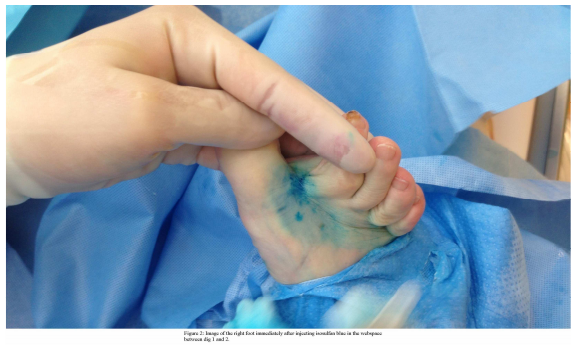
Some studies using isosulfan blue in the operative treatment of lymphoceles are reported. Stadelmann et al (2002) treated 19 lymphoceles in 17 different high risk patients, concluding that the use of isosulfan blue for identifying leaking lymphatic channels is successful. Of these 17 patients, none of the patients had an EVAR procedure; whereas three patients had an aortofemoral bypass. In all 19 surgically treated lymphoceles two wound abscesses and one superficial haematoma were reported, whereas no recurrence of the lymphocele was seen. In the study of Stadelmann isosulfan, blue dye was circumferentially injected into the distal extremity at the level of the ankle; the leg was then massaged and elevated to speed the migration of the dye. After an average follow up of 18,8 months all patients had a very faint residual blue hue at the injection site. For this reason we think it is more patient friendly to inject the dye in the webspace between digitus 1 and 2 of the foot.
In a study of Pagni et al (2009), two patients with persisting lymphoceles after non-surgical treatment were surgically treated. Isosulfan blue was used intraoperatively to map the lymphatic leakage. Complete resolution of the lymphocele occurred after ligation of the open lymphatics. As in our case they injected isosulfan blue intradermally in the webspace of the ipsilateral foot.
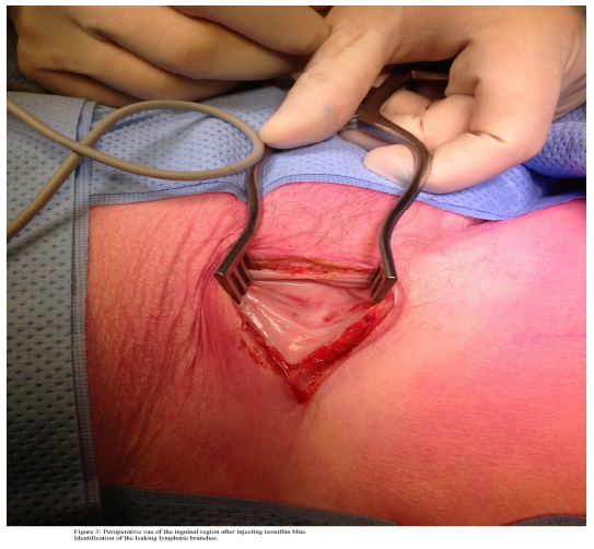
The standard surgical approach for lymfocele is excision of the lymfocele. Isosulfan blue can assists in identifying the leaking branches and facilitates in making the right excision, after identifying leaking lymphatics, ligation is possible.
Conclusion
We report a case of successful surgical treatment of a symptomatic groin lymphocele after EVAR. Isosulfan blue dye was helpful in identifying the lymphatic leakage.
Acknowledgements
The authors declare that they have no conflict of interest.
References
1. Berk, D.R., Johnson D.L., Uzieblo A., Kiernan M. and Swetter S.M. (2005) “Sentinel lymph node biopsy for cutaneous melanoma: the Stanford experience, 1997-2004.” Arch Dermatol, 141(8): p. 1016-22.
Publisher – Google Scholar
2. Bezu C., Johnson D.L., Uzieblo A., Kiernan M. and Swetter S.M. (2011) “Anaphylactic response to blue dye during sentinel lymph node biopsy.” Surg Oncol, 20(1): p. e55-9.
3. Choudhrie A.V., Kumar S., Gnanaraj L., Devasia A., Chacko N. and Kekre N.S. (2012) “Symptomatic lymphocoeles post renal transplant.” Saudi J Kidney Dis Transpl, 23(6): p. 1162-8.
Google Scholar
4. Hudack, S.S. and McMaster P.D., (1933) “The Lymphatic Participation in Human Cutaneous Phenomena : A Study of the Minute Lymphatics of the Living Skin.” J Exp Med, 57(5): p. 751-74.
Publisher – Google Scholar
5. Orvieto, M.A., Coelho R.F., Chauhan S., Palmer K.J., Rocco B. and Patel V.R. (2011) “Incidence of lymphoceles after robot-assisted pelvic lymph node dissection.” BJU Int, 108(7): p. 1185-90.
Publisher – Google Scholar
6. Pagni, R., Mariani C., Minervini A., Morelli A., Giannarini G. and Morelli G. (2009) “Treatment with intraoperative Patent Blue V dye of refractory lymphocele after inguinal lymphadenectomy for squamous cell penile carcinoma.” Urology, 74(3): p. 688-90.
Google Scholar
7. Pittaluga, P. and Chastanet S. (2012) “Lymphatic complications after varicose veins surgery: risk factors and how to avoid them.” Phlebology, 27 Suppl 1: p. 139-42.
Publisher – Google Scholar
8. Porcellini, M., Iandoli R., Spinetti F., Bracale U. and Di Lella D. (2002) “Lymphoceles complicating arterial reconstructions of the lower limbs: outpatient conservative management.” J Cardiovasc Surg (Torino), 43(2): p. 217-21.
Publisher – Google Scholar
9. Sansone, F., del Ponte S., Zingarelli E. and Casabona R. (2011) “The ‘packing of the groin’ technique: an innovative approach for groin lymphocele.” Interact Cardiovasc Thorac Surg, 13(4): p. 367-9.
Publisher – Google Scholar
10. Shermak, M.A., Yee K., Wong L., Jones C.E. and Wong J. (2005) “Surgical management of groin lymphatic complications after arterial bypass surgery.” Plast Reconstr Surg, 115(7): p. 1954-62.
Publisher – Google Scholar
11. Stadelmann, W.K. and Tobin G.R. (2002) “Successful treatment of 19 consecutive groin lymphoceles with the assistance of intraoperative lymphatic mapping.” Plast Reconstr Surg, 109(4): p. 1274-80.
Publisher – Google Scholar
12. Viehl, C.T., Guller U., Cecini R., Langer I., Ochsner A. and Terracciano L. (2012) “Sentinel lymph node procedure leads to upstaging of patients with resectable colon cancer: results of the Swiss prospective, multicenter study sentinel lymph node procedure in colon cancer.” Ann Surg Oncol, 19(6): p. 1959-65.
Publisher – Google Scholar
13. York, J.W., Johnson B.L., Cicchillo M., Taylor S.M., Cull D.L. and Kalbaugh C. (2013) “Aortobiiliac bypass to the distal external iliac artery versus aortobifemoral bypass: a matched cohort study.” Am Surg, 79(1): p. 61-6.
Google Scholar